Classification and General Characters

Digestive system of Pila :

Phylum - Mollusca - Soft bodied animal presence of hard calcareous
shell
Class - Gastropoda Foot in front of alimentary
canal
Subclass - Prosobranchiata Presence of gills or
ctinidium
Sab-order - Taenioglossa Ctinidium act as respiratory organ
Genus
- Pila
Species
- globosa

Characters
Pila
globosa is commonly known as apple snail it is Yoni valve freshwater mollusc it
is herbivorous and adapted for amphibious life in water and on land.
- The body the soft and protected by thick lemon yellow brownish or blackish calcareous shell.
- The shell is elongated hollow cone, spiral coilled circular around central axis called as collumela.
- The single revolution of sale around the central axis is called as whorl.
- There are usually 6 and half whorl. the top of a shell is icalled as Apex and the largest whorl close to the mouth is called as body whorl.
- The shell is marked by Mini lines of growth the vertical lines on shell are called varices (single-varix).
- The sexes are separate.
- The body whorl open outside by wide opening called as mouth, situated on ventral side, covered by a lid or operculum
- The region between two whorl is called suture.
Digestive system of Pila :
The digestive system consists of a long curved tube
extending from the mouth to the anus. It is broadly differentiated into
1.
Fore gut,
2.
Mid gut
3.
Hind gut

Fore gut:
The fore gut is ectodermal in origin and consists of
1.
Mouth
2.
Buccal mass
3.
Oesophagus.
Mouth: is a
median vertical slit situated at the anterior end of the snout that leads into
a thick walled muscular buccal mass.
The buccal mass encloses a cavity known as buccal
cavity which contains two jaws and a radula. At the entrance of the mouth is a
pair of chitinous jaws. The jaws bear
numerous small and two or three large teeth.
The buccal cavity shows chitinous ribbon shaped organ called radula.
Posteriorly, it is enclosed in a sac called the radular sac.
The radula bears numerous backwardly directed horny
teeth arranged in transverse rows. In Pila
each transverse row possesses seven teeth, a median rachidian teeth, on its
either side one lateral teeth and two marginal teeth, giving the formula 2, 1,
1, 1, 2
The teeth are
pointed with sharp edges and are used like a saw for cutting the food. The
radula is moved forward and backward, up and down with the help of protractor
and retractor muscles and act as a rasping organ.
A pair of salivary glands are present on the sides
of the buccal mass and their ducts open into the buccal cavity. The secretions
of the salivary glands contain mucus and an enzyme which digests starch. The
mucus lubricates the radula and helps in the transport of food.
Dorsally, the buccal mass leads into a long narrow oesophagus.
Just below the salivary glands, near the
origin of the oesophagus are a pair of round oesophageal pouches, probably
secrete digestive enzymes. Mid gut:
Mid gut is endodermal in origin
and consists of stomach and intestine. Oesophagus opens into the stomach. It is
red in color. The stomach consists of two parts:
1. Cardiac chamber and
2. Tubular
pyloric chamber.
The oesophagus opens into the cardiac chamber. The
cardiac chamber constitutes the main part of the stomach and possesses
longitudinal folds on its inner surface while the pyloric chamber has
transverse folds on its inner surface.
The intestine is long and forms 2 ½-3 coils. A
short, round blind pouch called the caecum arises from the pyloric chamber and
opens at the junction of the stomach and the intestine. Lying in the visceral
mass is the digestive gland (often referred to as the hepatopancreas or the
liver) which is dirty brown in color
The digestive gland contains three types of cells:
1. Secretory cells: they secrete cellulose digesting
enzymes.
2. Resorptive cells: they digest proteins
intracellularly.
3. Lime cells: they store calcium phosphate.
Hind gut:
The posterior part of the intestine is straight and
thick walled and is called the rectum. It terminates in an anus which is
situated near the mouth on the right side of the head The hindgut is ectodermal
in origin.
Nervous System of Pila Globosa:
The nervous system of Pila globosa is ganglonated type, consists of paired and unpaired ganglia with their commissures and connectives. The commissures are the connections between similar ganglia, while connectives are the nerves which connect two dissimilar ganglia.

Osphradium of Pila
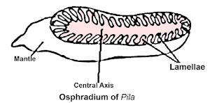
Location : It is a single, somewhat elongated structure suspend from the roof of mantle cavity close to the the left nuchal lobe or inhalant siphon.
Structure It is small elongated oval structure it is broader in in middle and at the right end it is bluntly rounded oval structure the left side is is somewhat pointed it consists of 22 to 28 thick fleshy roughly triangular leaflets. On median or central axis each leaflet is attached to the mantal wall bye its broad base and the central axis by it's smaller inner side
Osphradium is supplied by nerve from the left plural ganglion.
Function : it helps in to check the physico-chemical nature of water. that is it check the quality of water.
Radula of Pila


Location :
1. The radula is present in bucal cavity placed on a pair of radular cartilages operated by large radular muscles,
Structure :
2. Numerous transverse rows of teeth are present on the radular ribbon.
3. The teeth in a transverse row are one median or rachidian, one lateral and two marginal,
4. the radular formula being 2. 1. 1. 1. 2.
5. Distally, the radular teeth bear broad cutting edge
Function
The teeth are pointed with sharp edges and are used like a saw for cutting the food.
The radula is moved forward and backward, up and down with the help of protractor and retractor muscles and act as a rasping organ.
Nervous System of Pila Globosa:
The nervous system of Pila globosa is ganglonated type, consists of paired and unpaired ganglia with their commissures and connectives. The commissures are the connections between similar ganglia, while connectives are the nerves which connect two dissimilar ganglia.
The
paired ganglia of Pila are
1 Cerebral
ganglia
2 Buccal
ganglia
3 Pleural
ganglia
4 Pedal
ganglia
5 Visceral
ganglia,
Unpaired
ganglia are
1.
Supra-intestinal ganglia
2.
Infra-intestinal ganglia

1.
Cerebral ganglia:
These
are two triangular cerebral ganglia, present on dorslo-lateral side above the
buccal mass. These ganglia are connected
to each other by transverse connection called commissure above the buccal mass.
Cerebral ganglion are also connected with the buccal ganglion of its side
through a connection called cerebro-buccal connective. Cerebral ganglia aslo connected with pleural
and pedal ganglia by thick band-shaped cerebro-pleural and cerebro-pedal
connectives of the corresponding side.
Cerebral
ganglion gives off several nerves anteriorly to the skin of snout, the tentacle,
the buccal mass, the eye and the statocyst.
2.
Buccal Ganglia:
Buccal
ganglia are present at the junction of the buccal mass and oesophagus in the
form of pair. These ganglia are connected to each other by a transverse connection
called buccal commissure. They are also connected to the cerebral ganglia by a
cerebro-buccal connective on each side. The buccal ganglia supplies nerves to
the buccal mass, radular sac, salivary glands, oesophagus and the oesophageal
pouches.
3.
Pleuro-pedal ganglionic Mass:
The
Pleuro-pedal ganglionic Mass is formed by the fusion of the pleural and
pedal ganglia of each side lies just below the buccal mass. In a pleuro- pedal
ganglionic mass, the pleural ganglion is placed towards the outer side and the
pedal ganglion to the inner side. The pleuro-pedal ganglionic mass is connected
to the cerebral ganglion of its side by a connection called cerebro-pleural
connective and cerebro-pedal connective, respectively. While the two pedal
ganglia are connected to each other by two pedal commissures. To the right
pleuro-pedal ganglionic mass, infra-intestinal ganglion is fused. A delicate
loop-like infra-intestinal nerve behind
the pedal commissure, connects the pleural ganglia of both the sides.
4.
Supra-intestinal ganglion:
The
supra-intestinal ganglion is situated behind the pleuro-pedal mass of the left
side. Supra-intestinal ganglion is connected with the pleuro-pedal ganglionic mass
by a connective, called zygoneury. It gives thin supra-intestinal nerve on
inner side which joins to the right pleural ganglion.
The
supra-intestinal ganglion is also connected
to the visceral ganglion with visceral connective
5.
Visceral ganglion:
The
visceral ganglion is formed by the fusion of two spindle-shaped ganglionic
masses. The visceral ganglion lies near the base of the visceral mass close to
the anterior lobe of the digestive gland. The visceral ganglion is connected
with the supra-intestinal ganglion by a delicate supra-intestinal or left
visceral connective. It is also connected with the right pleural and
infra-intestinal ganglion through the infra-intestinal or the right visceral
connective.
Osphradium of Pila

Location : It is a single, somewhat elongated structure suspend from the roof of mantle cavity close to the the left nuchal lobe or inhalant siphon.
Structure It is small elongated oval structure it is broader in in middle and at the right end it is bluntly rounded oval structure the left side is is somewhat pointed it consists of 22 to 28 thick fleshy roughly triangular leaflets. On median or central axis each leaflet is attached to the mantal wall bye its broad base and the central axis by it's smaller inner side
Osphradium is supplied by nerve from the left plural ganglion.
Function : it helps in to check the physico-chemical nature of water. that is it check the quality of water.
Radula of Pila


1. The radula is present in bucal cavity placed on a pair of radular cartilages operated by large radular muscles,
Structure :
2. Numerous transverse rows of teeth are present on the radular ribbon.
3. The teeth in a transverse row are one median or rachidian, one lateral and two marginal,
4. the radular formula being 2. 1. 1. 1. 2.
5. Distally, the radular teeth bear broad cutting edge
Function
The teeth are pointed with sharp edges and are used like a saw for cutting the food.
The radula is moved forward and backward, up and down with the help of protractor and retractor muscles and act as a rasping organ.


